 A 10-month old white child was admitted for evaluation of an enlarged abdomen, splenomegaly, and developmental delay.
The child had a normal gestation and birth weight. He had a right hydrocele at birth and rapid scrotal enlargement at age 3 months that led to repair of a right inguinal hernia.
Physical examination revealed a large, somewhat lethargic child with unusual facies (Figure 1). Height, 32 inches (greater than the 97th percentile for age, 50th percentile for 18 months); weight, 24.5 lb (greater than the 97th percentile for age; 50th percentile for 16 months); and head circumference, 18.5 inches (50th percentile for 12 months). He had a small anterior fontanelle with prominent forehead, mild hypertelorism, a shallow nasal bridge with clear nasal mucus discharge, and a short neck.
The child's hands had a spadelike configuration, with ulnar deviation of the wrists.
Neuromuscular examination revealed normal muscle strength, tone, and deep tendon reflexes. Cranial nerve function was intact.
The liver edge was palpable 3 to 4 cm below the right costal margin.
Genital examination revealed a left scrotal mass with bilaterally descended testes and a small external urethral meatus.
Scattered maculopapular lesions were present over the patient's scalp, face, and neck. There was no lymphadenopathy.
The hematocrit was 38%; hemoglobin, 12.6 g/dL. The white blood cell count was normal, with granulation of the leukocytes on peripheral smear. Bone marrow analysis showed metachromatic granulation of histiocytes and leukocyte precursors. Levels of cholesterol, calcium, phosphorus, and total bilirubin were normal, as were results of tests of liver functions, routine karyotyping, electrocardiography, and skeletal survey radiography.
A family history revealed several maternal uncles with short stature; they had large heads, protruding abdomens, limited joint extension, hearing loss, poor vision, hernias, and cardiac abnormalities (Figure 2). A maternal great-uncle also had similar symptoms.

To what genetic disorder does this profile point?
(Answer and discussion on next page)
ANSWER: Hunter Syndrome
Mucopolysaccharide storage disease with X-linked recessive inheritance is typical of mucopolysaccharidosis (MPS) type II (Hunter syndrome).1 This disorder is now amenable to enzyme therapy.
MPS type II is caused by a deficiency of iduronate 2-sulfatase (I2S), a lysosomal enzyme involved in the breakdown of mucopolysaccharides (or glycosaminoglycans [GAGs]).1,2 With insufficient I2S, partially degraded GAGs accumulate in the brain, bones, heart, lungs, and visceral organs.
Because of maternal enzymes, an affected child is usually healthy at birth. Thereafter, gradual GAG storage causes:
•A developmental plateau and regression.
•Macrocephaly with potential hydrocephalus.
•A coarsened facial appearance (from thickened subcutaneous tissue).
•Visceral enlargement with hepatosplenomegaly (manifested by a protuberant abdomen).
•Skeletal changes, with metacarpal and vertebral processes evident on radiographs.
•Scoliosis/gibbus with joint contractures.
Early growth acceleration with later short stature is common. Without therapy, mental deterioration is relentless and culminates in a vegetative state and death.
GENETICSHunter syndrome is distinguished from other types of MPS by its X-linked rather than autosomal recessive inheritance. That is, it is transmitted on the female X chromosome from mother to child.

 Figure 3 shows this patient's maternal male relatives, with coarsened facial appearance and skeletal changes that have been likened to those of gargoyle statues. The pedigree provides strong evidence of X-linked recessive inheritance by male-only affliction, lack of male-to-male transmission, and linkage of affected males through females (female carriers) (see Figure 2). The 2 X chromosomes in females allow them to "carry" an abnormal allele on 1 X chromosome that is compensated for by a normal allele on the companion X. In future pregnancies, carrier females have a 1:2 or 50% risk that any son will be affected with Hunter syndrome and a 1:2 or 50% risk that any daughter will be a carrier.
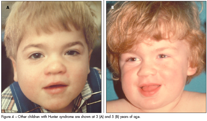
Hunter syndrome occurs in all ethnic groups. The incidence is slightly higher among certain Israeli Jewish populations, in whom the incidence is 1 in 65,000 to 1 in 132,000 births.1,2 Other male patients with the coarsened facies of Hunter syndrome are shown in Figure 4.
 MPS DISEASE SUBTYPES
Hunter syndrome occurs as a type A (MPS II A) that presents during childhood and a type B (MPS II B) that may not cause symptoms until adulthood. Those with type B have similar physical findings once the disease manifests; however, the severe skeletal problems and/or neurodegeneration seen in type A disease do not develop.
The facial features of patients with Hunter syndrome1 are common to several other MPS types. These include:
•The very severe Hurler syndrome (MPS I H).
•The less severe Maroteaux-Lamy syndrome (MPS VI).
•Sanfilippo syndrome, which has a more gradual onset with severe behavior abnormalities (MPS III--4 types).
•The rare Sly syndrome (MPS VII).
•The type IV Morquio syndrome, which has less striking facial changes and more severe skeletal deformities of the chest and ribs than Hurler or Hunter syndromes.1,2
The skeletal changes, joint contractures, and valvular stenoses that accrue from mucopolysaccharide storage clearly illustrate that these syndromes are connective tissue and neurological disorders. Pulmonary changes with thick secretions, restrictive lung disease, and aspiration/pneumonias in the vegetative state are the usual causes of death.
Other signs and symptoms of MPS include macrocephaly with hydrocephalus, short neck, broad chest, protuberant abdomen with hepatosplenomegaly and umbilical/inguinal hernias, vision impairment with cloudy corneas and/or atypical retinitis pigmentosa, and sensorineural or conductive hearing loss. (Patients with Hunter syndrome lack corneal clouding but tend to have cream-colored papules over the upper back.) Inspection of leukocytes on peripheral smears or in bone marrow aspirates will show granules caused by GAGs stored in lysosomes.
OLIGOSACCHARIDOSESEnzyme deficiencies in the degradation pathways for GAGs and complex lipids account for a related category of disease called oligosaccharidoses (OS). These disorders were called mucolipidoses; however, the name has changed to the more accurate chemical term. OS disorders include GM1 gangliosidosis, I (inclusion)-cell disease, sialidoses, and others that may be more severe than MPS. Patients with OS may show signs at birth and display features of lipidoses, such as the retinal cherry red spot seen in Tay-Sachs disease.2
MPS and OS are distinguished from other storage diseases by urine testing (increased amounts of GAGs or oligosaccharides). The diagnosis is made by assay of white cells (anticoagulated blood--green-top tube) that can be ordered as a lysosomal enzyme panel.1,2 Assay of the I2S enzyme deficient in Hunter syndrome is performed at relatively few laboratories. DNA diagnosis should soon supplant enzyme assays.
If enzyme assays or DNA tests confirm that a child has an MPS or OS, prenatal testing can be considered should the child's mother again become pregnant. Strategies for prenatal diagnosis include preimplantation diagnosis with blastocyst selection after in vitro fertilization, chorionic villus sampling (at 10 to 12 weeks' gestation), or amniocentesis (at 15 to 18 weeks' gestation).
The patient in our case presented before such assays were available. Nevertheless, the elevated urine glucopolysaccharide level and the patient's severely afflicted maternal relatives support a diagnosis of Hunter syndrome (MPS II A).
MANAGEMENTClinical management of patients with Hunter syndrome or related MPS/OS is directed toward anticipating and ameliorating symptoms. Early childhood intervention for developmental delays and joint limitations, careful monitoring for hydrocephalus or vision/hearing changes, nasal saline rinses and aggressive respiratory treatment, alertness for hernias or bowel problems, and orthopedic care for bone changes are paramount. Initial genetic and later supportive counseling for a terminal disease is important. Management checklists have been published.3
Newer therapies include bone marrow transplantation with release of normal I2S enzyme that improves somatic features of Hunter syndrome. Results with regard to crossing the blood-brain barrier and improved neurologic status are variable. In a study of 10 patients, only 3 survived 7 years after bone marrow transplant. Physical and mental disabilities had progressed in 2 of those 3 patients; intellectual development had remained normal in the third patient, who had mild physical disability.4
Recombinant DNA technology is now used for I2S enzyme production.5 Suitable cultured cells are infected with a recombinant virus (vector) engineered to express the gene that encodes I2S enzyme. After cell growth with suitable I2S synthesis, the enzyme is purified, dried, and stored in vials for reconstitution and intravenous administration. Like other lysosomal enzymes, the I2S is targeted to and taken up by lysosomes via a specific mannose 6-phosphate signal site. Once in the lysosomes, the enzyme degrades accumulated GAGs and improves somatic symptoms. Whether I2S and other recombinant enzymes (including laronidase [Aldurazyme] for Hurler syndrome) can effectively be transferred across the blood-brain barrier is still in question. 
The biosynthetic I2S enzyme idursulfase (Elaprase) has recently been approved by the FDA. Intravenous administration of this enzyme improved joint mobility and diminished organ size during clinical trials in 96 persons with Hunter syndrome.6
The availability of enzyme therapy and possible limited access to accumulated GAGs in the brain emphasizes the importance of early recognition and diagnosis of Hunter syndrome and other MPS disorders.


